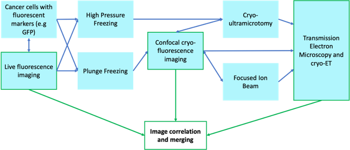
Cryo-Correlative Light & Electron Microscopy (Cryo-CLEM) is an innovative pipeline that enables specific targeting of macromolecular structures for structural biological characterisation in situ, i.e. in the contact of intact cells. The equipment listed on this page consists of various vitrification and imaging equipment that is essential in this pipeline in order to enable Cryo-Electron Tomography (Cryo-ET), or 3D cryo-electrom microscopy of vitreous samples. The Workflow enables the investigation of the complex changes in macromolecular structure that are intrinsic to the mechanism of action of molecular machines and are not easily reconstituted in vitro. Below is a schematic of example workflows:
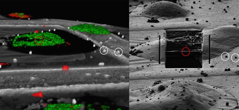
Picture of fluorescence signal overlaid to ion beam image during Cryo-FIB-milling for ET. Picture generated from Leica.
Equipment & Technology:
• EM GP2 Plunge Freezer – for rapid cryo-fixation and vitrification of thin samples on grids.
• EM ICE High Pressure Freezer – for vitrification of thicker samples (e.g. organoids, tissue).
• Ultracut + FC7 Ultramicrotome for CEMOVIS (Cryo Electron Microscopy of Vitreous Sections)
• Stellaris 5 Cryo Confocal – to record and transfer co-ordinates of suitably preserved cells and identify intracellular macromolecular targets for cryo-FIB Milling and cryo-ET
| Hours | Location |
|
Monday - Friday 9:30 - 17:30h |
Chester Beatty Laboratories |
1.
| Name | Role | Phone | Location | |
|---|---|---|---|---|
| Kai Betteridge |
LM Facility Manager
|
kai.betteridge@icr.ac.uk
|
.-N112.1
|
|
| Teige Matthews-Palmer |
EM Facility Manager
|
Teige.Matthews-Palmer@icr.ac.uk
|
5C9
|
|
| Queenie Lai |
Microscopist
|
Queenie.lai@icr.ac.uk
|
.-1N12.1
|
|
| Ross Scrimgeour |
Microscopist
|
Ross.scrimgeour@icr.ac.uk
|
.-1N12.1
|
|
| Matthew Jessop |
CryoET and CLEM Scientist
|
matthew.jessop@icr.ac.uk
|
5C9
|